About the program
Three-quarters of cancer patients are over the age of 60, but certain types of cancer occur in younger people and even children.
Good results in cancer treatment are achieved only with early recognition, when there are no symptoms yet. Early pre-clinical diagnosis of cancer is possible through whole-body examination using MRI and the latest PET-CT imaging available to modern medicine. These two studies make it possible to detect foci as small as 2 mm, their metastases to organs, bones, lymph nodes, to establish the degree of spread of the tumor, and, accordingly, the stage of the disease with an already diagnosed disease. Both methods of diagnostics are necessary for the early detection of relapses of oncological diseases. None of these studies has practically no contraindications.
Oncological program
- clinical blood test with leukocyte formula
- biochemical blood test (liver and kidney tests, carbohydrate and lipid metabolism, proteins and protein fractions, electrolytes, blood clotting parameters)
- blood test for thyroid hormones (to assess its function)
- blood test for pancreatic hormones (for the diagnosis of diabetes mellitus at the preclinical stage, as well as diseases of the pancreas)
- for men: test for prostate specific antigen (PSA)secreted by cells of the prostate gland (to diagnose prostate cancer and monitor the treatment of existing prostate disease)
What is this?
Method for studying the heart muscle by recording bioelectrical signals of the heart at rest from the body surface.
For what?
For the diagnosis of cardiac arrhythmias and other pathologies of the heart (cicatricial changes, ischemia, etc.).
What is this?
The method of diagnosing the vessels of the fundus using a special diagnostic
installation that allows you to fix the arteriovenous ratio of retinal vessels, and
also changes in the fundus in digital form.
For what?
The state of the fundus vessels allows you to assess the risk of stroke
brain.
What is this?
A method for studying the function of external respiration, including the measurement of lung volumes, as well as speed indicators of pulmonary ventilation.
For what?
Used to assess the functional state of the respiratory system.
What is this?
A combination of ultrasound diagnostics and color coding of moving blood with a two-dimensional image of the lumen and vessel walls.
For what?
Ultrasound and DS of extracranial and intracranial vessels helps to identify obstructions to blood flow, arterial and venous aneurysms, occlusion of the vessels of the base of the brain and vertebral artery, to assess the degree of stenosis of the carotid arteries and atherosclerotic lesions, to determine the spasm of the cerebral arteries.
UZDG and DS neck vessels is carried out to assess the condition of the walls of blood vessels and analyze blood flow. Thus, the thickness of the internal vascular wall is determined, circulatory disorders in the carotid arteries, zones of thrombosis or vascular compression, as well as the presence of atherosclerotic deposits are detected.
What is this?
A non-invasive method for obtaining images of internal organs using ultrasound.
For what?
Ultrasound of the thyroid gland and soft tissues of the neck allows you to reliably judge the state and size of the thyroid gland and identify areas of heterogeneity in its tissues. The results of the examination indicate the presence or absence of nodular pathology of the thyroid gland, cysts, inflammation.
What is this?
Non-invasive study by ultrasound diagnostics of all structures of the heart in the process of its work.
For what?
Using this method, the wall thickness, the volume of the heart cavities, the condition and functioning of the valvular apparatus, the contractile function of the heart muscle are determined, intracardiac hemodynamics, blood flow parameters in the heart chambers are examined, intracardiac thrombi, neoplasms, heart aneurysm, heart defects are detected.
What is this?
Method for obtaining a three-dimensional image of the main and peripheral vessels with
intravenous administration of contrast agents.
For what?
MRA is used to detect congenital abnormalities of the blood vessels,
atherosclerotic deposits, areas of narrowing and blockage of arteries, consequences
stroke, as well as the diagnosis and subsequent choice of methods for treating diseases
blood vessels.
What is this?
Imaging method of internal organs and soft tissues based on exposure
magnetic field on the human body at the molecular level. Computer fixes
response of the body to this effect and interprets it in the form of a clear
layer-by-layer image of internal organs and soft tissues of the body.
For what?
MRI is performed to diagnose diseases at an early stage. Advantage
of this method of examination lies in the fact that the patient is not exposed to
X-ray exposure, while the quality of diagnosis is not reduced.
MRI of the whole body makes it possible to detect hidden foci of diseases in the body
different (often unexpected) localizations, as well as to conduct an accurate analysis of the likely
painful changes in the head, neck, spine, chest and
abdominal cavity, pelvis and hip joints. The method is indispensable for express
diagnostics as part of a preventive examination of the body.
What is this?
Imaging method of internal organs and soft tissues based on exposure
magnetic field on the human body at the molecular level. Computer fixes
response of the body to this effect and interprets it in the form of a clear
layer-by-layer image of internal organs and soft tissues of the body.
For what?
MRI is performed to diagnose diseases at an early stage. Advantage
of this method of examination lies in the fact that the patient is not exposed to
X-ray exposure, while the quality of diagnosis is not reduced.
MRI of the head includes examination of the diencephalon and medulla oblongata,
pituitary gland, epiphysis, cerebellum, cerebral hemispheres, its membranes,
vessels, ventricles, cerebrospinal fluid pathways, as well as the skull, ENT organs and
contents of the eye sockets, This method is especially effective in identifying
benign and malignant tumors, metastases, foci of infection,
vascular disorders, atherosclerotic plaques, aneurysms, cysts. In addition, with his
with the help of detecting diseases in the area of the eye orbits, paranasal sinuses,
nasopharynx.
What is this?
Imaging method of internal organs and soft tissues based on exposure
magnetic field on the human body at the molecular level. Computer fixes
response of the body to this effect and interprets it in the form of a clear
layer-by-layer image of internal organs and soft tissues of the body.
For what?
MRI is performed to diagnose diseases at an early stage. Advantage
of this method of examination lies in the fact that the patient is not exposed to
X-ray exposure, while the quality of diagnosis is not reduced.
MRI of the mammary glands reveals pathological formations in the breast, their size and
localization, helps to distinguish benign tumors from malignant ones, and
also determines the presence of cysts and hematomas in the breast tissue. MRI is the most
effective method in the early diagnosis of breast cancer and in the detection of
recurrence of neoplasms.
What is this?
The method of layer-by-layer examination of internal organs and tissues, based on
using the capabilities of x-ray technology and a computer. Computer
the program analyzes the degree of absorption of x-rays and reconstructs
examined organs and tissues in a three-dimensional image.
For what?
Chest CT can diagnose diseases of the lungs, heart, esophagus,
large blood vessels (aorta, pulmonary artery), soft tissue damage
the central part of the chest. This survey can detect
infectious diseases (tuberculosis, pneumonia, pleurisy, etc.), lung cancer or
metastases from other organs, emphysema and bronchiectasis, pulmonary embolism and aneurysm
aorta, mediastinal pathology.
CT of the heart is used to determine the presence and extent of atherosclerosis,
cholesterol deposits in the coronary vessels of the heart and heart attack risk assessment
myocardium.
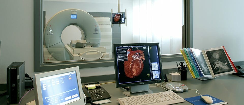
What is this?
The combination of two modern methods of examination - positron emission
tomography (PET) and computed tomography (CT). For this survey
The patient is injected intravenously with a radioactively labeled substance, which
accumulates and differently distributed in the cells of the body, revealing foci
pathologies, and computed tomography helps to accurately localize them.
For what?
PET-CT is the most advanced method of diagnosing cancer, able to detect
malignant tumors at the so-called "zero" stage, when there are still no
any symptoms. PET-CT is also used to determine the stage of cancer and
detection of latent metastases. In addition, this study is used in cardiology,
neurology and other areas of medicine.
Duration: 1 day.