If the suspicion of testicular cancer is confirmed, then the diseased testicle must be completely removed by surgery (orchiectomy).
After that, follow-up studies are carried out. They should, first of all, show whether the lymph nodes are affected or metastases have appeared in other organs. The goal of treatment is to remove the cancerous tissue completely or destroy it in order to achieve a cure.
The first step in the treatment of testicular cancer is usually the surgical removal of the affected testicle (orchiectomy). Only in advanced cases of the disease can the removal of the affected testicle be postponed, and chemotherapy is first performed. What further treatment methods are carried out after an orchiectomy depends on the type of tumor (seminoma or non-seminoma) and how far the disease has gone at the time of diagnosis.
Further treatment with semin
For patients in whom it is established that there is no damage to the lymph nodes and the spread of the tumor to other organs (N0M0), there are three options for further treatment. With the expectant strategy (so-called Wait—and—see—strategy) further therapy is not carried out. Points to watch out for: It is known that, despite negative diagnostic results, approximately 20% patients have tiny affected lymph nodes in the retroperitoneum. Thus, these 20% patients will have a recurrence of the disease in the future if no further treatment is carried out immediately after the operation. Since, with timely detection of larger findings, the chances of recovery are no less than with immediate therapy, the patient can choose this strategy. But control examinations must be carried out very carefully and at regular intervals. If the patient decides in favor of immediate treatment to reduce the risk of 20% recurrence, then he has two options: either retroperitoneal irradiation along the abdominal aorta or chemotherapy with a well-tolerated drug. Irradiation is carried out 5 times a week and ends after 2 weeks. Chemotherapy is prescribed on an outpatient basis and consists of 1 or 2 injections of the drug. The chances of recovery in all three variants are almost 100%. For the patient to choose one of them, a detailed conversation with the attending physician is required. If there are metastases in the retroperitoneal space in the lymph nodes with a maximum diameter of up to 5 cm (N1 - N2), then chemotherapy is carried out, as well as in cases of lesions of larger lymph nodes (N3) or metastases in other organs (M+).
Further treatment with nonsemin
With non-seminomas without data on the spread of the tumor to the lymph nodes and other organs, it is also possible to conduct an expectant strategy (Wait—and—see—strategy). The risk of recurrence can be assessed using prognostic factors. The risk is especially high if, when examining the tissues of the removed testicle under a microscope, it is clear that the tumor cells have penetrated into the blood vessels of the testicle (vascular invasion). In this case, one should not take a wait-and-see attitude, since the risk of metastasis is approximately 50%. As a preventive treatment, the following methods are available: removal of certain regions of lymph nodes in the retroperitoneal space (diagnostic retroperitoneal lymphadenectomy) or chemotherapy with one or two drugs. The treatment is carried out in a hospital for 6 weeks, and the patient during this period of time is in the hospital only on certain days. There is damage to the blood vessels - chemotherapy, as a systemic therapy, is the most reliable method. Recent studies have shown that the appointment of even one cycle of chemotherapy is very effective. Patients with existing lymph node metastases and/or metastases to other organs are now recommended chemotherapy. With lymph nodes up to 2 cm in size in the retroperitoneal space and negative tumor markers in the blood, it makes sense to surgically remove them, or, as an option, first conduct a control and, in case of a further increase in their size, surgical removal or chemotherapy. The number of cycles of chemotherapy is guided by the prevalence of lymph node damage, which organs are affected and how high the rate of tumor markers is. A tumor larger than 1 cm found in the body after chemotherapy must be removed promptly. The individual therapies are presented in more detail below.
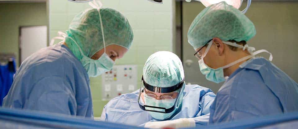
Operation
Testicle removal (orchiectomy)
An orchiectomy is the first step in the treatment of testicular cancer. In this case, the affected testicle is first exposed. As a rule, the operating doctor immediately sees whether it is a malignant tumor or not. If this is not clear in some rare cases, then a piece of tissue is taken and examined under a microscope during the operation. The suspicion of testicular cancer is confirmed, the diseased testicle with the epididymis and the vas deferens is removed. This operation is called an orchiectomy. The operation is relatively uncomplicated and safe. During the operation, a tissue sample (biopsy) can be taken from a healthy testicle to rule out a precancerous stage (testicular intraepithelial neoplasia, TIN) that could develop there. If such a precancerous stage is detected, the testicle is subjected to radiation therapy. Irradiation causes, as a consequence, a disturbance in the production of semen and thus the infertility of the patient. If you want to have children, there is an alternative: wait first, and start treatment only when a malignant tumor develops. This can take years under certain circumstances.
The removal of one testicle (semicastration) does not affect either sexuality or potency, or the possibility of conceiving children. The healthy testicle of the opposite side takes over the function of the removed testicle. However, in 50% testicular cancer patients, for unknown reasons, seed production in the healthy testis is limited. Therefore, after removal of the affected testicle, it makes sense to examine the semen quality of the remaining testicle by taking a semen sample before subsequent therapy. If indeed there is limited semen production, then sperm can be collected and frozen for artificial insemination later. This is important, since further therapy leads to damage, even with the passage of 2-3 cycles of chemotherapy and to a temporary one. For cosmetic reasons, the removed testicle can, if desired, be replaced with a prosthesis that looks and feels like a healthy testicle.
Removal of lymph nodes (retroperitoneal lymphadenectomy)
During this operation, the lymph nodes in the retroperitoneal space are removed. The extent of the operation depends on the stage of the tumor. If the lymph nodes on computed tomography are without features (N0), then the operation is performed on a modified field, which means the removal of lymph nodes only in certain areas. If lymph node involvement is confirmed (N1-2), then the operating field is expanded. In each case, doctors make an effort to keep nerve fibers in the surgical field that are important for the release of semen (ejaculation). If they are damaged, sperm during orgasm is not ejected through the urethra outward, but in the opposite direction (retrograde) into the bladder (retrograde ejaculation). The patient's ability to conceive naturally is lost. But spermatozoa can be isolated from urine and used for artificial insemination. In most cases, in the early stages of the disease, nerve-sparing surgery is possible on a limited operating field so that normal sperm ejection is preserved. In any case, potency, libido and the ability to orgasm are preserved.
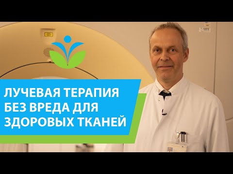
Radiation therapy
Radiation therapy uses radioactive radiation to destroy cancer cells. It can be used in the treatment of seminomas. But the condition for its use is the disease at an early stage, which means the absence or presence of only small metastases in the lymph nodes, and the absence of metastases in other organs (N0-2). The region of the posterior wall of the abdomen is irradiated to the left and to the right of the abdominal aorta. This should prevent the development of metastases in the lymph nodes (N0), or their metastases (N1-2) are completely destroyed. If in this area metastases to the lymph nodes have already been detected (N1-2), then additionally on the affected side, the pelvic area is irradiated along the large vessels of the pelvis. As a rule, irradiation is carried out on an outpatient basis. It is important to weigh the potential long-term consequences of radiotherapy against morbidity with alternative therapies. For example, the use of radiation therapy for clinical stage 1 seminoma (tumor extension limited to the testis) has been heavily criticized, as the study showed that 14% patients should expect a second malignant tumor after 18 years.
Radiation therapy is the treatment of choice if a biopsy of the opposite testicle reveals an early form of cancer (testicular intraepithelial neoplasia, TIN)
Chemotherapy
Chemotherapy aims to destroy cancer cells throughout the body with cell-growth-inhibiting drugs (cytostatics). Cytostatics work well against cells that grow very quickly, this property is characteristic of cancer cells. In testicular cancer, chemotherapy is prescribed, as a rule, in cases where the disease has spread throughout the body. In seminomas, chemotherapy is the method of choice in advanced stages of the disease, when there are large metastases in the lymph nodes (N3) or metastases formed in other organs (M1). For non-seminomas, chemotherapy can be given already in the early stages of the disease. In this case, it is a complementary (adjuvant) therapy immediately after the removal of the testicle (N0), or after removal of the affected lymph nodes of the retroperitoneal space (N1-2). Patients in advanced stages, which means with large lymph node metastases (greater than 5 cm) or organ metastases (N3, M1), always receive primary chemotherapy. As a rule, with this therapy, metastases in the lymph nodes and organs can be destroyed. Subsequently, in case of nonseminomas, the remnants of the tumor in the abdominal cavity are removed, since in approximately 50% patients, despite chemotherapy, live tumor cells remain. Approximately 20% of these are considered malignant, but 30% living cells that do not show the typical microscopic signs of malignancy can grow back and must therefore be removed. With seminoma after chemotherapy, the remnants of the tumor are only observed at first, since, as a rule, we are talking about dead tissue. Using positron emission tomography, it should be determined whether there is simply scar tissue or living tissue with carbohydrate metabolism. If carbohydrate metabolism is detected, then the mass should be removed. If new growth occurs at follow-up, then further treatment should be considered. At far advanced stages of the tumor, as a rule, 4 courses (cycles) of chemotherapy are performed. Usually the same medicines are used as in the early stages, but they can also be changed depending on the size of the tumor. The cycle of chemotherapy takes 21 days, and medications are administered only on certain days, between which the patient can recover at home.
High dose chemotherapy
For patients with very advanced disease, such as liver, brain, or skeletal metastases (poor prognosis group IGCCCG) is currently being tested for the value of high-dose chemotherapy. Apply high doses of various cytostatics. Since intensive treatment destroys not only tumor cells, but also hematopoietic cells of the bone marrow, the patient's stem cells are taken before high-dose chemotherapy and returned to the body after treatment (blood stem cell transplantation). Chemotherapy at high doses is currently carried out only in special centers in the framework of scientific research.
Treatment of relapses
In the case of a return of the disease (relapse), an attempt is made to achieve recovery again by surgical removal of the tumor or by new chemotherapy, more often with a subsequent operation. The duration and intensity of treatment are focused on the size and location of the recurrence.


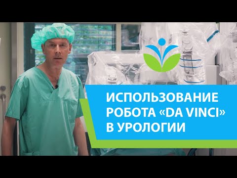
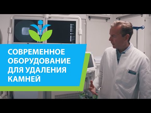
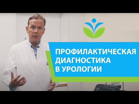
Video


Request appointment
Useful links


Photo gallery