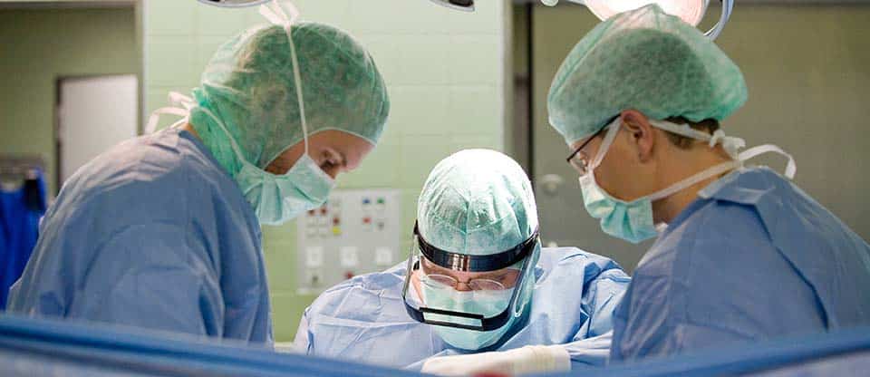
Thanks to a clear organization and continuity in the diagnosis and surgical treatment of breast diseases, the necessary operation is carried out as soon as possible after the diagnosis is made. Diagnostic recognition of the tumor by mammography and/or ultrasound examination until the most accurate histological confirmation of the tumor is completed is about 18 hours. The use of an operative method of cancer treatment allows surgeons to save the mammary glands in 70% cases. This became possible with the use of plastic reconstructive surgery methods, while using the body's own tissues (tissues of the breast itself and adjacent tissues). In the event that the breast still needs to be removed, the patients are offered the possibility of plastic breast reconstruction.
Techniques that ensure breast preservation
After organ-preserving operations for breast cancer, it is mandatory to carry out postoperative irradiation of the gland according to the scheme at certain intervals.
The removed lymph nodes, the tumor and the nearby tissue surrounding the tumor are subject to a thorough histopathological examination to establish the fact of a tumor lesion of the lymph nodes. When examined under a luminescent microscope, cells of the sentinel lymph nodes retain a specific color, which makes it possible to detect even the smallest fragments of cancer cells. The results of histopathological examination of malignant neoplasms are important for the diagnosis and treatment of malignant tumors in the mammary gland. This is the highest authority, which gives the final conclusion about the type of tumor, the need for surgery on a (still) healthy organ, and much more. Decisive for the diagnosis are biochemical studies of the surface parts of tumor cells. One of the possibilities of biochemical studies is to establish the sensitivity of the tumor to hormones. The condition of the lymph nodes is an essential criterion for determining the stage of the tumor process, and staging serves to describe the disease. After determining the stage of the disease and prognostic factors, doctors have the opportunity to draw up a treatment plan, which is then discussed in detail with the patient.
Removing nodes
Under general anesthesia or local anesthesia, the skin is cut directly over the tumor. Then the tumor is carefully removed with a margin within healthy tissues. The marking of the tumor allows histologists to know exactly how the removed tumor was located in the gland. The defect in the area of remote nodes, in most cases, is small, and there is no need for its plastic reconstruction. As a rule, it is not externally noticeable that part of the breast tissue has been removed. A cosmetic suture is applied to the incision site, which after a while is barely noticeable. The described method of node removal is suitable only for small tumors and the absence of the need for plastic reconstruction of the postoperative defect.
Partial removal of glandular breast tissue
The surgical method by which the removal of the glandular tissue of the mammary gland is carried out is similar to the method described above for the removal of nodular neoplasms. Difference: with partial removal of glandular tissue, a larger volume of tissue is removed. The muscle tendons under the tumor are always removed, and sometimes the area of skin above the tumor. The resulting extensive tissue defect, in most cases, requires plastic reconstruction of the breast. Based on the size of the mammary gland, a postoperative defect can be eliminated using tissues from the same operated mammary gland by turning or moving the tissue.
Often, after surgery, a significant cosmetic defect occurs on the mammary gland affected by the tumor process due to the difference in the size of both glands. In such a situation, in order to eliminate the defect, the patient is offered to reduce a healthy breast. After receiving detailed information about the method, the patient must decide on the possibility of performing the operation herself. Only the voluntary consent of the patient is the basis for this type of reconstruction. If the patient considers it necessary to reconstruct the part of the mammary gland remaining after surgical treatment to the shape and size of a healthy gland, plasty is performed by transferring tissue from the patient's back to replace the postoperative defect. And the doctor informs the patient in detail about this possibility of plastic replacement in a mandatory conversation before the operation.
The removed lymph nodes, the tumor and the nearby tissue surrounding the tumor are subject to a thorough histopathological examination to establish the fact of a tumor lesion of the lymph nodes. When examined under a luminescent microscope, cells of the sentinel lymph nodes retain a specific color, which makes it possible to detect even the smallest fragments of cancer cells. The results of histopathological examination of malignant neoplasms are important for the diagnosis and treatment of malignant tumors in the mammary gland. This is the highest authority, which gives the final conclusion about the type of tumor, the need for surgery on a (still) healthy organ, and much more. Decisive for the diagnosis are biochemical studies of the surface parts of tumor cells. One of the possibilities of biochemical studies is to establish the sensitivity of the tumor to hormones. The condition of the lymph nodes is an essential criterion for determining the stage of the tumor process, and staging serves to describe the disease. After determining the stage of the disease and prognostic factors, doctors have the opportunity to draw up a treatment plan, which is then discussed in detail with the patient.
Complete exfoliation of glandular breast tissue
With the help of an electric knife, scissors or a scalpel, surgeons manage to completely peel off the glandular tissue of the breast from the skin. Replacement of a tissue defect is carried out by means of prostheses or own tissues.
It is necessary to pay attention to the fact that irradiation of the prosthesis after the operation does not make sense, since complications may occur in 50% cases. The prosthesis can be used only if it is known for sure that postoperative irradiation will not be carried out, or the prosthesis will be thoroughly covered with muscle tissue as a result of the operation. Of particular importance in such cases is the close interaction of surgeons-mammologists and radiologists.
Simultaneous removal of the breast nipple
All mammary excretory ducts of the mammary glands gather at the top of each mammary gland, forming the mammary nipples. If the primary localization of cancer is the milk ducts, then the operation requires the removal of the nipple. Sometimes it happens that the surgeon during the operation cannot recognize whether there is a tumor lesion of the milk ducts. In this case, the histological conclusion is of particular importance. With an affirmative answer of the histologists, an additional minor operation to remove the nipple is indicated. Sometimes nipple removal can change the whole concept of therapy. In this case, the attending physician explains in detail to the patient the reasons for the change in treatment tactics.
The removed nipple can then be reconstructed using various techniques. If a young woman has undergone surgery to remove the nipple, unfortunately, she will not be able to breastfeed her child.
The removed lymph nodes, the tumor and the nearby tissue surrounding the tumor are subject to a thorough histopathological examination to establish the fact of a tumor lesion of the lymph nodes. When examined under a luminescent microscope, cells of the sentinel lymph nodes retain a specific color, which makes it possible to detect even the smallest fragments of cancer cells. The results of histopathological examination of malignant neoplasms are important for the diagnosis and treatment of malignant tumors in the mammary gland. This is the highest authority, which gives the final conclusion about the type of tumor, the need for surgery on a (still) healthy organ, and much more. Decisive for the diagnosis are biochemical studies of the surface parts of tumor cells. One of the possibilities of biochemical studies is to establish the sensitivity of the tumor to hormones. The condition of the lymph nodes is an essential criterion for determining the stage of the tumor process, and staging serves to describe the disease. After determining the stage of the disease and prognostic factors, doctors have the opportunity to draw up a treatment plan, which is then discussed in detail with the patient.
Breast removal
In about 30% cases of cancer, complete removal of the breast is inevitable. The radical method significantly increases the chances of a complete cure. Often there are cases when women, in order to avoid postoperative radiation, decide on a complete removal of the breast. This is especially true for older women. After removal of the mammary gland, a barely noticeable oblique scar on the chest remains.
In some cases, it is possible to save part of the tissue in the area of the midline of the mammary gland, which makes it possible for a woman to create a beautiful decollete zone. And the lateral areas of the mammary gland are replaced in the bra with silicone pads. Another option: silicone pads are glued to the skin. In addition, at the individual request of patients, operative-reconstructive plastic surgery is used to restore the shape and size of the breast.
Removal of lymph nodes
Classic radical removal
Radical removal of lymph nodes is always an integral part of breast removal surgery. Cells of malignant tumors grow into lymphatic and blood vessels, can be torn off and transported by blood and lymph flow to other organs and tissues. Taking into account the structure of the lymphatic system, its protective function in the body, the ways of metastasis of malignant cells, during a radical operation for breast carcinoma for more than a hundred years, the standard technique is considered expedient to simultaneously remove regional lymph nodes.
There are various methods for this. For example, the classic removal of at least 10 lymph nodes, or the removal of several "sentinel" lymph nodes (Sentil limph node) - the lymph nodes closest to the mammary gland. Most often, they are the first organs where the tumor metastasizes. Histological examination of the removed lymph nodes allows surgeons to draw conclusions about the presence of cancer cells in the lymph nodes of the axillary region.
According to the classical surgical technique, from the surgical wound on the mammary gland or from an additional incision in the armpit, adipose tissue is removed with lymph nodes of 1 and 2 levels of metastasis, sometimes even 3 levels of metastasis. Lymph from the mammary gland flows into the sentinel nodes (Sentinel lymph node), then malignant cells from the tumor focus through the lymphatic ducts can penetrate into the axillary lymph nodes. To determine the possible spread of malignant cells from the sentinel nodes in the armpit, there is a special technique. The technique consists in the fact that a radioactive substance is injected into a place near the tumor with a syringe in a very small dose. Malignant cells, represented by a radioactive substance, reach the following lymph nodes with the lymph flow and mark them.
The operating surgeon using a special apparatus (think of a small Geiger counter) can determine whether there is metastasis of malignant cells.
In the narrow anatomical space of the armpit, numerous clusters of nerves and blood vessels are closely located. Therefore, the classical extended removal of lymph nodes in the armpit leads to negative consequences, which for many patients are aggravating. Approximately half of all patients who underwent removal of lymph nodes by the classical method for many years after the operation complain of limited movement in the shoulder girdle, a feeling of numbness on the inner surface of the shoulder, extended scars in the armpit, swelling of the arm (lymphatic edema).
Dissection of sentinel lymph nodes (Sentinel node)
This surgical method is not yet a common standard! This is a very gentle method of removing lymph nodes and is performed only in a few clinics. To perform an operation using this method, a long-term experience of the surgeon is required with very good collaboration with radiation medicine doctors. During the operation, radiologists introduce a substance labeled with radioactive isotopes into the patient's body. This technique can be compared with the well-known examination of the thyroid gland (thyroid scintigraphy). For two years in the Breast Center of the St. Antonius Clinic, operations using the method of preparation of sentinel lymph nodes have been the standard. The head physician of the clinic has many years of experience (based on previous work in specialized clinics around the world) in the surgical technique of dissecting sentinel lymph nodes.
Determination of the stage of the disease - prognostic factors
The removed lymph nodes, the tumor and the nearby tissue surrounding the tumor are subject to a thorough histopathological examination to establish the fact of a tumor lesion of the lymph nodes. When examined under a luminescent microscope, cells of the sentinel lymph nodes retain a specific color, which makes it possible to detect even the smallest fragments of cancer cells. The results of histopathological examination of malignant neoplasms are important for the diagnosis and treatment of malignant tumors in the mammary gland. This is the highest authority, which gives the final conclusion about the type of tumor, the need for surgery on a (still) healthy organ, and much more. Decisive for the diagnosis are biochemical studies of the surface parts of tumor cells. One of the possibilities of biochemical studies is to establish the sensitivity of the tumor to hormones. The condition of the lymph nodes is an essential criterion for determining the stage of the tumor process, and staging serves to describe the disease. After determining the stage of the disease and prognostic factors, doctors have the opportunity to draw up a treatment plan, which is then discussed in detail with the patient.
The removed lymph nodes, the tumor and the nearby tissue surrounding the tumor are subject to a thorough histopathological examination to establish the fact of a tumor lesion of the lymph nodes. When examined under a luminescent microscope, cells of the sentinel lymph nodes retain a specific color, which makes it possible to detect even the smallest fragments of cancer cells. The results of histopathological examination of malignant neoplasms are important for the diagnosis and treatment of malignant tumors in the mammary gland. This is the highest authority, which gives the final conclusion about the type of tumor, the need for surgery on a (still) healthy organ, and much more. Decisive for the diagnosis are biochemical studies of the surface parts of tumor cells. One of the possibilities of biochemical studies is to establish the sensitivity of the tumor to hormones. The condition of the lymph nodes is an essential criterion for determining the stage of the tumor process, and staging serves to describe the disease. After determining the stage of the disease and prognostic factors, doctors have the opportunity to draw up a treatment plan, which is then discussed in detail with the patient.
Video
Request appointment
Useful links


