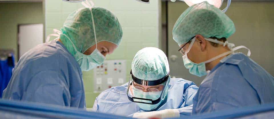
If you suspect uterine cancer (endometrial carcinoma, cancer of the uterine body), the doctor prescribes the necessary examinations. With their help, you can find out whether we are really talking about a tumor process, and if so, what type of tumor is present and how far the disease has gone. Decisive for clarifying the diagnosis is the histological microscopic examination of tissue samples. This study is necessary in any case.
With a refined diagnosis of endometrial cancer, further examinations are connected, which should show how far the tumor has spread, whether the lymph nodes are affected, and whether metastases have appeared in other areas of the body. However, there are no technical means that could replace surgical staging (determining the extent of a tumor disease through surgical intervention) for uterine cancer. If the tumor is inoperable due to concomitant diseases, then MRI may be useful for planning the therapy of a local neoplasm, and CT for assessing the state of the lymph nodes, respectively.
Only when all the necessary examinations are completed, the doctor and the patient jointly decide which treatment measures are best suited.
Conversation with the doctor (anamnesis)
Before prescribing an examination and treatment for uterine cancer in Germany, the doctor asks in detail about the presence of complaints and since when they appeared. In addition, during the collection of a family history, it finds out possible risk factors.
Gynecological examination with examination in the mirrors
During this examination, the doctor examines the walls of the vagina and cervix with a special tool in order to determine where, respectively, from the uterus, whether the bleeding came from, and whether the tumor has grown outside the uterus. In addition, swabs may be taken for microscopic examination (cytology).
Clinical examination (palpation)
In conclusion, a palpation examination of the uterus is performed. At the same time, the doctor palpates the body of the uterus through the abdominal wall, through the vagina - the cervix, uterine os, as well as the tissues surrounding the uterus for possible changes. Additionally, tissues surrounding the uterus are felt through the rectum. By palpation examination, the doctor can already obtain important information about the nature and extent of the disease.
Transvaginal ultrasound (sonography)
With this method, the doctor can evaluate the lining of the uterus. Here you can determine the thickness of the mucosa. In a woman with post-menopausal bleeding, mucosal thickness greater than 5 mm is suspicious for cancer. In addition, it is often possible to determine how deeply the malignant process has penetrated into the muscular layer of the uterus and whether the fallopian tubes and ovaries are affected. If uterine cancer is suspected, a transvaginal ultrasound is always performed.
Hysteroscopy
During hysteroscopy, a probe with a small video camera with a built-in light source is inserted into the uterine cavity through the vagina. For a better view of the mucous membrane, the uterine cavity can be washed with liquid. Tissue samples are taken from suspicious areas with small forceps, which are later examined under a microscope. Hysteroscopy is usually combined with curettage of the uterine cavity.
Curettage of the uterine lining (fractionated curettage)
Separate histological examination under a microscope of a tissue sample from the cervix and uterine cavity mucosa is currently the most reliable method for diagnosing endometrial cancer. To obtain a sample, a careful separate curettage of the uterine cavity through the vagina is carried out. In this case, fragments of the mucous membrane of the cervical canal and the mucous membrane of the body of the uterus are taken and examined (fractionated curettage). The mucous membrane is restored over time under the influence of hormones. Sample collection can be combined with hysteroscopy. At the same time, it is possible to purposefully take samples from suspicious areas before scraping. By combining both methods, malignant tumors are diagnosed with great reliability.
Cystoscopy and rectoscopy
Cystoscopy and rectoscopy are performed when there is a suspicion that the tumor has already reached the bladder or rectum. In this case, the probe is passed through the urethra into the bladder and through the anus into the rectum. At the same time, the doctor can examine the walls of the bladder and intestines and take tissue samples from suspicious areas, which will then be examined histologically under a microscope.
Computed tomography (CT) and magnetic resonance imaging (MRI)
CT and MRI are used to determine the extent of a tumor in the abdominal cavity. In this case, metastases or enlarged lymph nodes can be determined. Nevertheless, the exact scale of the prevalence of the tumor process can only be determined within the framework of the operation. A CT scan is ordered to evaluate the lymph nodes.
MRI also allows you to present the body in the form of sections. However, the examination is carried out not under X-rays, but in a magnetic field. This is intended to examine the structure and relationship of tissues relative to each other.
Laboratory research
The blood test provides information about the general condition of the patient, as well as the function of individual organs, such as the kidneys and liver. The results of the research are important for assessing the situation, taking into account the upcoming operation. In addition, the so-called "oncomarkers" are determined. In this case, we are talking about substances that are produced by tumor cells and can be found in the blood. Some types of endometrial cancer sometimes produce tumor markers that can be detected in the blood. They are called CA 125 (cancer antigen 125) and CEA (carcinoembryonic antigen). However, tumor markers are not found in all patients with uterine cancer and may be higher than normal in healthy people.
Therefore, for diagnosis, they are only relevant for certain (rare) forms. First of all, they serve to control the dynamics of the disease, if they were elevated at the beginning of the disease.


Head of the Center for Operative Gynecology
Head of the Regional Center for Pelvic Floor Surgery


Video


Request appointment
Useful links


Photo gallery