
If bowel cancer is suspected, your doctor may do an endoscopic examination of your bowel (colonoscopy). If there is a suspicion of bowel cancer, various examinations may be prescribed. With their help, it is possible to establish whether there is actually a tumor disease, and if so, how far it has gone.
The most important examination methods for detecting a tumor:
- Palpation (with the introduction of fingers into the rectum)
- Test for occult blood in stool
- Rectoscopy (endoscopy with a rigid endoscope of the rectum to a depth of 20 cm)
- Sigmoidoscopy (flexible endoscopy of part of the large intestine)
- Colonoscopy (endoscopy with a bendable endoscope of the entire large intestine). Colonoscopy is the most important and informative study for the diagnosis of intestinal tumors. If cancer is suspected, tissue samples (biopsy) will be taken. These samples are examined under a microscope by a specialist for the presence of tumor cells. Only after this study can we say for sure whether there is a tumor disease or not. Colonoscopy is the method of choice. In addition, colonoscopy is included in the complex of studies used for the early diagnosis of bowel cancer.
In the case of an oncological diagnosis, further examinations are carried out to find out how much the tumor has spread to neighboring tissues and organs.
These include:
- Ultrasonography (sonography/endosonography). In order to determine how much a malignant tumor (rectal carcinoma) has spread, an ultrasound examination is performed by introducing a special probe into the intestine. Assess the degree of its distribution in the intestinal wall and the condition of the nearby lymphatic glands. Of particular value is endosonography for planning surgery. Based on its results, the doctor determines whether the rectal sphincter will be preserved or whether an intestinal stoma is required. Endosonography also helps in deciding whether to prescribe radiation therapy or radiation plus chemotherapy.
- Computed tomography (CT). This X-ray method, in which the human body is translucent layer by layer, allows you to accurately determine whether the cancer has gone beyond the walls of the intestine and whether it has affected neighboring organs or lymph nodes (metastasis). And when planning radiation therapy, computed tomography provides assistance.
- Magnetic resonance imaging (MRI). For colorectal cancer, magnetic resonance imaging is used to assess the size and extent of the tumor. It is especially good to use MRI before surgery to assess the anatomy and extent of the tumor in relation to the rectal sphincter.
- Laboratory research. For the treatment of tumors of the large intestine and rectum, the so-called tumor markers are of primary importance. An elevated content of tumor markers in the blood may be an indication of a malignant tissue change. Unfortunately, tumor markers are not absolutely specific and can be elevated in healthy people. In addition, a negative result or a level of tumor markers within the normal range does not completely and reliably exclude cancer, since not all cancer patients have an elevated level of tumor markers. The analysis for tumor markers is especially suitable for monitoring the course of the disease after surgery, if their level was elevated before that. Therefore, analysis for tumor markers is used as part of patient monitoring for timely recognition of relapse. For intestinal tumors, the most important is the determination of the oncomarker CEA (cancer-embryonic antigen).
Only after all examinations are completed, it is possible to plan the necessary treatment.

Doctor of Medical Sciences
Head of the Clinic for General, Visceral and Minimally Invasive Surgery
Head of the Clinic for General, Visceral and Minimally Invasive Surgery

Professor, MD, PhD
Head of the Clinic for General, Visceral, Thoracic and Endocrine Surgery
Head of the Clinic for General, Visceral, Thoracic and Endocrine Surgery


Video
Precise mathematical calculations for the most effective radiation therapy.
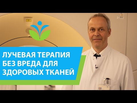
Radiation therapy without harm to healthy tissue? In our clinic it is possible!
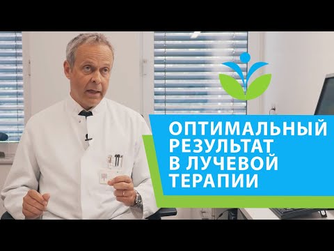
Guaranteeing optimal results in oncology - an interdisciplinary approach in radiotherapy.
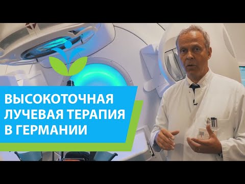
A complete cure for cancer is possible thanks to the high precision of radiation therapy.
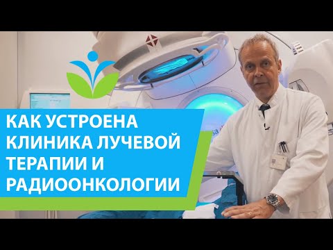
How does a radiation therapy and radiation oncology clinic work in Germany? (12+)
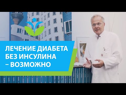
“Treating diabetes without insulin is possible!” - Professor Martin proves.
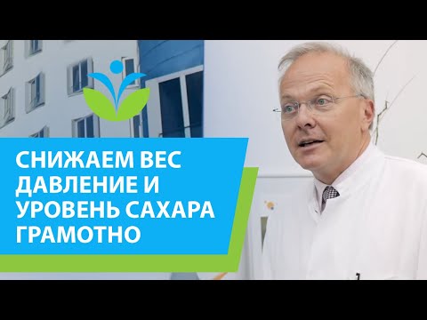
We reduce weight, pressure, sugar levels competently - a program awarded with an international scientific award
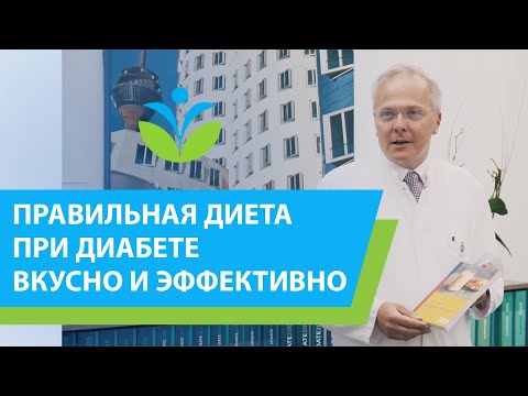
The right diet for diabetes is tasty, simple, effective and without the need for medications (12+).
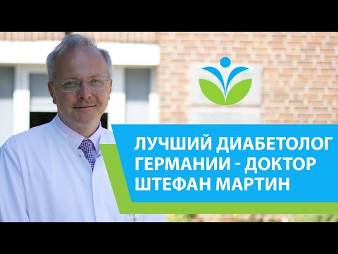
The best diabetes doctor in Germany about the innovative program together with Deutsche Klinik Allianz.
Combined oncology treatment at the Bethesda Clinic is comprehensive and effective.
A unique center for thyroid surgery under the guidance of a leading specialist in Europe.
Why the clinic takes on complex medical cases and successfully handles them - says Dr. Zimon.
The best operating ophthalmologist in Germany about his experience of working with foreign patients.
Ophthalmologist Dr. Thomalla is an innovator who performed the world's first cornea transplant (6+).

About the treatment of reflux - the best surgeon in Germany Konstantinos Tsarras
Request appointment
Useful links


Photo gallery